Tập tin:Brain - Lobes.png
Không có độ phân giải cao hơn.
Brain_-_Lobes.png (701×487 điểm ảnh, kích thước tập tin: 360 kB, kiểu MIME: image/png)
| Miêu tả |
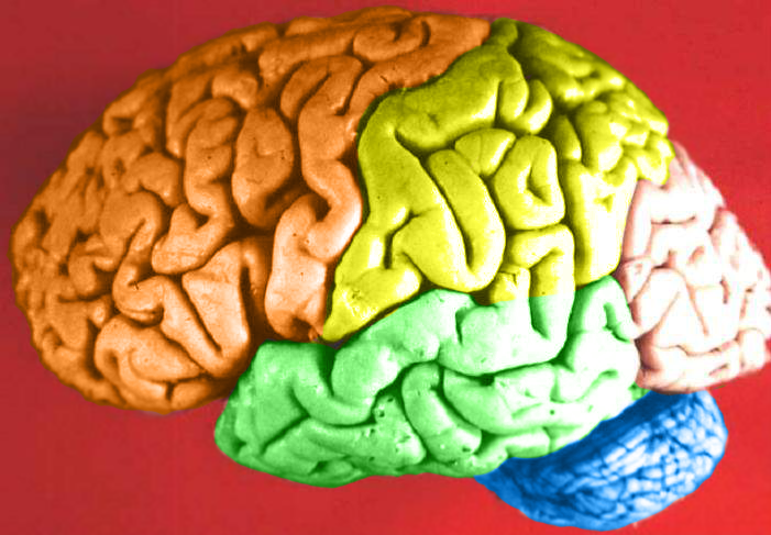
Human brain lateral view - Lobes
|
| Ngày | (UTC) |
| Nguồn gốc | Human_brain_lateral_view_description.JPG |
| Tác giả | Dep't. of Cellular Biology & Anatomy, Louisiana State University Health Sciences Center Shreveport |
| Giấy phép (Dùng lại tập tin) |
CC-BY |
| Phiên bản khác |
| Đây là một ảnh đã được chỉnh sửa, có nghĩa là nó đã được chỉnh sửa kỹ thuật số so với phiên bản gốc. Các chỉnh sửa được thực hiện bao gồm: Hemispheres in color.. Có thể xem phiên bản gốc tại đây: Human brain lateral view description.JPG. Các chỉnh sửa được thực hiện bởi DavoO.
|
Giấy phép
Tôi, người giữ bản quyền tác phẩm này, từ đây phát hành nó theo giấy phép sau:
Tập tin này được phát hành theo Giấy phép Creative Commons Ghi công 2.5 Chung.
- Bạn được phép:
- chia sẻ – sao chép, phân phối và chuyển giao tác phẩm
- pha trộn – để chuyển thể tác phẩm
- Theo các điều kiện sau:
- ghi công – Bạn phải ghi lại tác giả và nguồn, liên kết đến giấy phép, và các thay đổi đã được thực hiện, nếu có. Bạn có thể làm các điều trên bằng bất kỳ cách hợp lý nào, miễn sao không ám chỉ rằng người cho giấy phép ủng hộ bạn hay việc sử dụng của bạn.
The following refers to the original source file, not this derivative version.
Tập tin này, vốn được đăng tải tại https://web.archive.org/web/20110514023714/http://www.healcentral.org/healapp/showMetadata?metadataId=40566, đã được người duyệt hình Avenue kiểm tra vào ngày 1 tháng 11 năm 2013 và xác nhận rằng nó đã được phát hành dưới giấy phép tương ứng trong ngày hôm đó.
|
Nhật trình tải lên đầu tiên
This image is a derivative work of the following images:
- File:Human_brain_lateral_view_description.JPG licensed with Cc-by-2.5
- 2006-06-20T13:58:22Z Patho 701x487 (50176 Bytes) {{Information| |Description='''Human brain lateral view - Lobes''' # Lobus frontalis # Lobus parietalis # Lobus temporalis # Lobus occipitalis # Sulcus lateralis # Sulcus centralis # Sulcus parietooccipitalis # Incisura preo
- 2006-06-20T13:54:13Z Patho 701x487 (49891 Bytes) Auf eine alte Version zurückgesetzt
- 2006-06-20T13:51:38Z Patho 701x487 (50074 Bytes) {{Information| |Description='''Human brain lateral view - Lobes''' # Lobus frontalis # Lobus parietalis # Lobus temporalis # Lobus occipitalis # Sulcus lateralis # Sulcus centralis # Sulcus parietooccipitalis # Incisura preo
- 2006-06-20T13:28:44Z Patho 701x487 (49891 Bytes) {{Information| |Description='''Human brain lateral view''' # Lobus frontalis # Lobus parietalis # Lobus temporalis # Lobus occipitalis # sulcus lateralis # Sulcis centralis # Sulcus parietooccipitalis # Incisura preoccipital
Uploaded with derivativeFX
Lịch sử tập tin
Nhấn vào ngày/giờ để xem nội dung tập tin tại thời điểm đó.
| Ngày/Giờ | Hình xem trước | Kích cỡ | Thành viên | Miêu tả | |
|---|---|---|---|---|---|
| hiện tại | 05:11, ngày 24 tháng 2 năm 2009 | | 701×487 (360 kB) | wikimediacommons>DavoO | {{Information |Description='''Human brain lateral view - Lobes''' # Lobus frontalis # Lobus parietalis # Lobus temporalis # Lobus occipitalis # Sulcus lateralis # Sulcus centralis # Sulcus parietooccipitalis # Incisura preoccipitalis # Polus frontalis # |
Trang sử dụng tập tin
Trang sau sử dụng tập tin này: